Table of Contents
Foreign body removal from Eye
To understand the technique of foreign body removal, first of all, we should know what foreign body is? What types of foreign body usually enter in the eye? So, let discuss it.

What is a foreign body in the eye?
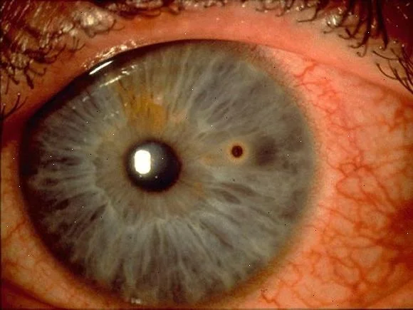
Foreign Body in the eye means any types of foreign things, Object, and material which come in contact with an eye in a different situation. The Corneal Foreign body and Conjunctival Foreign body are the common foreign body in the eyes.
Types of the foreign body:
Chemical (lime, H2SO4, HCL) in the eye, Dust, Sand, Iron particle, Insect wing etc.
Ocular Foreign Body Removal Technique:
There are various practices in ophthalmology to remove the foreign body from the eye.
Let’s discuss one by one:
- Irrigation of eye
- Finding the foreign body Position, location and depth and sweeping it.
- Removal of F.B. in OPD under Topical anaesthesia in slit lamp.
- Removal of a foreign body under local anaesthesia/ GA in an operating microscope.
1. Irrigation of eye:
Indications for irrigation of an eye are biohazard exposures, chemical irritants, acid or alkaline burns, and loosely attached foreign body removal. A Morgan lens attached to flowing normal saline can be inserted to assist in irrigation provided that the cornea is not Perforated or a foreign body is not well embedded into the cornea. Do not delay irrigation for a detailed history, vision testing, or examination. Ideally, the pH should be checked before irrigation as long as this does not delay irrigation. Litmus paper or urine dipstick can be used to check the pH.
Irrigation with 3-4 liters of normal saline may be adequate in acid exposure if pH is maintained at neutrality 30 minutes after completion of irrigation. In alkaline exposures, irrigation may be required for 24 hours. The safest method is a careful irrigation of the eye with a stream of sterile saline, directed from the sclera over the cornea. It is important to evert the lids and irrigate both superior and inferior fornices.
Credit: Pak Sang Lee, Community Eye Health Journal
2. Finding the F. B. Position, location and depth and then sweeping it
In the examination of the eye to check for a foreign body, it is important to view all the cornea, sclera and palpebral conjunctiva by everting the lid. Foreign bodies under the lid or on the sclera may be wiped away by a cotton-tipped applicator. Wetting the applicator will often enhance its effectiveness.
Sweeping the palpebral conjunctiva will often relieve the symptoms even if the foreign body is not directly seen.
 Karin Lecuona/Dept. of Ophthalmology University of Cape Town, CEH
3. Removal of F.B. in OPD under Topical anaesthesia in slit lamp/ Loop
A corneal/ scleral foreign body that is not deeply penetrating the cornea can be removed in the emergency room under slit lamp/ or using a loop.

4. Removal of a foreign body under local anaesthesia/ GA in operating microscope
But if the foreign body is very deep-seated and very difficult to remove in the emergency room i.e. in OPD. Then we should send the patient in OT and removal can be done under local anaesthesia with Operating microscope
Note: Always check the patient’s TETANUS immunization status.
If the foreign body is iron-based, follow up is needed because of a propensity to form a RUST RING.


