

Optic Disc Changes in Glaucoma : Diagnosing Glaucoma Case
Primary open angle glaucoma is a common eye disease characterized by loss of the axons of the retinal ganglion cells leading to progressive loss of vision. The site of damage to the axons is at the level of the lamina cribrosa in the optic nerve head. The mechanism of axonal loss is unknown but elevated intraocular pressure and age are the most common factors associated with the disease.
Previous studies in human glaucoma and in experimental glaucoma in monkeys have established a relationship between chronic elevation of intraocular pressure and remodelling of the optic nerve head tissues known clinically as cupping of the optic disc. This review focuses on the astrocytes, the major cell type in the optic nerve head. Astrocytes participate actively in the remodelling of neural tissues during development and in disease. In glaucomatous optic neuropathy, astrocytes play a major role in the remodeling of the extracellular matrix of the optic nerve head, synthesize growth factors and other cellular mediators that may affect directly, or indirectly, the axons of the retinal ganglion cells.
Due to the architecture of the lamina cribrosa, formed by the cells and the fibroelastic extracellular matrix, astrocytes may respond to changes in intraocular pressure in glaucoma, leading to some of the detrimental events that underlie axonal loss and retinal ganglion cell degeneration.
Neuroretinal Rim
Related Posts
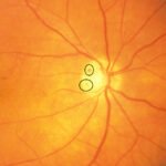
The area of the disc occupied by the retinal nerve fibre axons is referred to as the neuroretinal rim and it is changed in the appearance of this area that is the key![Neuroretinal Rim of optic disc]() to quantifying glaucomatous disc damage. When examining the neuroretinal rim the pattern of thickness and areas of focal thinning are the most important features.
to quantifying glaucomatous disc damage. When examining the neuroretinal rim the pattern of thickness and areas of focal thinning are the most important features.
 to quantifying glaucomatous disc damage. When examining the neuroretinal rim the pattern of thickness and areas of focal thinning are the most important features.
The ISNT rule is useful in evaluating whether thinning is physiological or pathological. A healthy disc tends to have its thickest portion of neuroretinal rim inferiorly, then superiorly, then nasally, with the thinnest portion being temporal.
ISNT rule
inferior![]()

superior
nasal
temporal
In fact, the temporal rim being the thinnest is probably the most important. However, it is also important to remember that some healthy discs will not obey the ISNT rule, and similarly some glaucomatous discs may obey the ISNT rule.
The ISNT guideline
No one has yet been able to elicit an absolute biomarker (be it physical or chemical) for glaucoma or the risk thereof. Maybe such a “God particle” for glaucoma just doesn’t exist. I agree with the aforementioned studies, but that doesn’t mean I don’t think about the ISNT rule when looking at an optic nerve. I would change it up a bit semantically, though, by calling it the “ISNT guideline” instead. I’m a fan of the ISNT rule (guideline) for several reasons. However, the biggest reason I think it has a place in contemporary eye care is the fact that it forces the clinician to think about the optic nerve head in terms other than simply a decimal point with a number after it. I’m not saying cup-to-disc ratios are not of the upmost importance with respect to glaucoma. I’m just saying that if someone were to describe an optic nerve head to me by saying, “point six,” I’d be on the edge of my seat waiting for more information.
So, even if the ISNT rule doesn’t prove to be true (in sensitivity or specificity) a lot of the time, it forces the clinician to really examine the entire tissue of the optic nerve head (any of which may be damaged early on in glaucoma). Further, looking past the cup-to-disc ratio gives the clinician the opportunity to look for other potential aspects of the glaucomatous optic nerve, such as the presence of retinal or optic nerve hemorrhages, areas of zone beta parapapillary atrophy, or retinal nerve fiber layer defects around the optic nerve.
I, for one, find the ISNT rule to be a useful tool of which to be mindful. It certainly isn’t intended to be taken as an absolute truth. It makes me stop and look a little more closely at optic nerves, however, and for that reason, I enjoy keeping the ISNT rule in mind.
Truly, the exciting and highly useful advancements in spectral domain optical coherence tomography (SD-OCT) have given us more ammunition in the battle to diagnose and treat glaucoma earlier, but close, careful, and direct stereoscopic evaluation of the optic nerve head, itself, must not become a lost art. A diagnosis of glaucoma starts at the optic nerve, and adages such as the ISNT rule remind us of this important point.ODT
Focal notching:
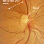
Focal notching is very characteristic of glaucoma and tends to be inferio- or superio- temporal. It can become a full-thickness notch with complete abscence of the rim. It may be very deep and it may be associated with peripapillary atrophy. Progression will cause this area to enlarge. Notches usually correspond to specific visual field defects so it is major sign of Glaucomatous Optic Disc changes,
Optic disc haemorrhage
Splinter haemorrhage (Disc Harmorrhage) can be seen in normals but are associated with the progression of the disease, particularly in NPG. particular seen in the infero-temporal region. It may precede optic disc change and field loss. Optic cup enlargement can progressive in advance of pallor.
Retinal nerve fibre layer (RNFL)defects
The retinal nerve fibre layer is the innermost layer of the eye and corresponds to the nerve fibres passing from the photoreceptors over the retina as they course towards the optic disc. In the same way the loss of these fibres causes the optic cup to expand, the loss of fibres can be evaluated by looking at the presence or absence of this layer of nerve fibres particularly in young people.
wedge defects-their presence is significant, their absence is of no significance.
- RNFL is seen with the green light on the slit lamp.
- RNFL can be photographed.
- RNFL analysers looking for loss of the retinal nerve fibre layer are available but are of limited use at present. They are very poor at screening for glaucoma but may be useful in monitoring ocular hypertension/early glaucoma for progression. Once there is moderate disease they are very poor at demonstrating subtle changes.
Peri-papillary atrophy

This is a feature of the normal aging process and of high myopia. It may however also be a feature of glaucoma.
In glaucoma patients, the extent of the atrophy has been shown to correlate with the severity of the disease and progression of disease, particularly in NTG.
In non-glaucoma PPA is most common temporally. In glaucoma, the area of PPA is usually in the sector with the rim loss.
In a senile sclerotic disc, it has been reported to be present for the full 360dgrees.
References
1. Morgan JE, Bourtsouki I, Rajkumar KN, et al. The accuracy of the inferior>superior>nasal>temporal neuroretinal rim area rule for diagnosing glaucomatous optic disc damage. Ophthalmology. 2012 Apr;119(4):723-30.
2. Hwang YH, Kim YY. Application of the ISNT rule to neuroretinal rim thickness determined using Cirrus HD optical coherence tomography. J Glaucoma. 2013 Nov 14.
3. Pogrebniak AE , Wehrung B, Pogrebniak KL, et al. Violation of the ISNT rule in nonglaucomatous pediatric optic disc cupping. Invest Ophthalmol Vis Sci. 2010 Feb;51(2):890-5.