

Lines in Ophthalmology simply means the lines seen on various parts of the eyes. Lines in eyes is important because of it may give hints of the past or recent eye health conditions. Here are some of the eponymous signs in the ophthalmology field.
Table of Contents
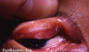
Arlt’s Line
Arlt line is a thick, dense, horizontal band of scarring near the lid margin. It is an important finding of an eye disease called trachoma where chronic inflammation occurs in tarsal conjunctiva. This Arlt’s line runs horizontally, parallel to an eyelid, this line is located at the junction of the anterior 1/3rd and posterior 2/3rd of the conjunctiva.
Ehrlich-Turck Line
This line is seen on the cornea. It is due to the linear deposition of keratic precipitates on the corneal endothelium. It is a vertical line seen on the Uveitis (Inflammation of uvea).
Ferry’s Line
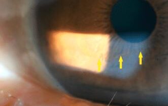
Ferry’s line is a subepithelial iron deposit at the corneal edge of a filtrating bleb after trabeculectomy.
Fingerprint lines
Fingerprint lines are also known as, map-dot-fingerprint dystrophy, epithelial basement membrane dystrophy. It is an unusual appearance in the cornea. This type of dystrophy seen in both eyes and usually affects adults between the ages of 40 and 70. This is usually asymptomatic which means the patient doesn’t have any symptoms.
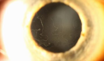
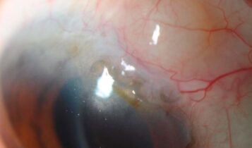
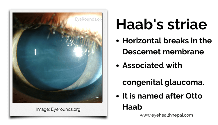
Haab striae (The Descemet Break)
Haab striae is corneal horizontal or concentric breaks in the Descemet’s membrane. It is Similar to posterior polymorphous dystrophy (PPMD). However on histopathology: the edge of Haab’s striae are thickened, curled, with the area between the edge being smooth and thin. This helps differentiate from PPMD. Descemet folds seen on congenital glaucoma or buphthalmos. Descemet’s breaks or from birth trauma tend to be vertical, while Descemet’s tears associated with congenital glaucoma tend to be horizontal or curvilinear.
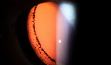
Hudson-Stahil Line
It is a normal and common finding in elderly patients. Hudson-Stahil Line is an iron deposition line in the corneal epithelium, it is commonly observed in the junction between the middle and lower third of the cornea. The thickness of this Hudson Stahl line is about 0.5 mm and its length is 1-2 mm. This line causes no symptoms or any clinical significance.
Khodadoust Line
Khodadoust Line or chronic focal transplant reaction is seen on the corneal transplant (Post keratoplasty) eyes. It is an ocular sign of corneal transplant surgery complications on the eye. This line is seen on the corneal endothelium due to mononuclear cells / keratic precipitates accumulating there.
This line is a sign of corneal graft endothelial cell rejection. If untreated, the line of WBCs will move across and damage the endothelial cells of the cornea within few days, so it needs immediate immunosuppression treatment to prevent further damage at an early
Krukenberg’s spindle
Krukenberg’s spindle is the name given to the pattern formed on the inner surface of the cornea by pigmented iris cells that are shed during the mechanical rubbing of the posterior pigment layer of the iris with the zonules that are deposited as a result of the currents of the aqueous humour. It occurs in association with “classic” or primary pigment dispersion syndrome (PDS) and pigmentary glaucoma.
LASIK Iron Line
Epithelial iron lines can be seen on some of the post-refractive surgical procedures. The tear film
distribution may be altered after LASIK surgery which allows some pooling centrally. This pooling can cause iron deposition in the central epithelium of the cornea.
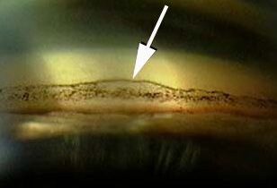
Paton’s Line
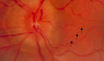
It is located at the optic disc (Retina). Paton’s line is a vertical, circumferential retinal fold especially temporal to the optic disc. This line indicates the optic disc (Optic nerve head) swelling (papilledema) It is mainly due to blockage of the axoplasmic transport at the lamina cribrosa and secondary causes is due to increased in Intracranial pressure (ICP).
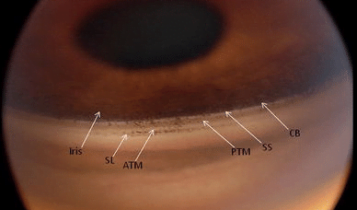
Sampaolesi line
The pigment found anterior to or along Schwalbe’s line is called Sampaolesi’s line. Although this line can be idiopathic, or sometimes this line indicate pseudoexfoliation (PXF) syndrome, pigment dispersion syndrome glaucoma etc. When Sampaolesi’s line is visible there may have hyperpigmentation throughout the angle, especially in the Trabecular Meshwork (TM).
Schwalbe’s Line
It is a gonioscopic view of the drainage angles. It is a termination of Descemet’s membrane of the cornea. This is a line where corneal scleral meshwork terminates anteriorly.
Stockers Line
Stocker’s line is a yellow or brown deposition in the corneal epithelium. It is Iron deposition that can be visible close to the leading edge of a pterygium. A vertical line at the head of the pterygium is known as the stockers line.
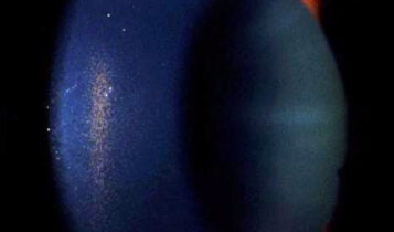
Vogt’s striae
Vogt’s striae occur at the central cornea in a patient having keratoconus. when applying pressure on the globe digitally while looking through the slit lamp, these striae in the deep cornea mostly disappears. This line is the characteristic of keratoconus. It is a vertical stromal/Descemet’s membrane line. It is stress lines due to stretching and thinning of the cornea. This is one of the important lines in ophthalmology.

White lines of Vogt
The white line of Vogt is the Sheathed or sclerosed vessels in lattice degeneration. Lattice is a degenerative disease of the peripheral retina in which the lattice lines are presents created by fibroid blood vessels. Lattice degeneration is present in roughly only 10% of lesions and most likely represent hyalinized blood vessels.
Zentmeyer line (Scheie’s Line)
Pigment accumulated at the zonular attachments to the lens Although some report it as pigment on the peripheral posterior lens capsule. Scheie’s line” is considered to be pathognomonic for pigment dispersion syndrome.
Four corneal iron lines
- Stocker line: Pterygium
- Fleischer line: Keratoconus
- Ferry line: Filtering bleb
- Hudson-Stähli line
These lines are clearly visible on slit lamp by using either cobalt blue or red-free filters without using fluorescein.
These above-mentioned lists of lines in ophthalmology important streaks and striae were seen in the various parts of eyes, these are the most used common lines, streak and striae. You can also easily differentiate between Vogt’s striae vs Haab’s striae ophthalmology lines by studying the above-mentioned details.
You can also like our other post on Rings in Ophthalmology.